

AI Brain Imaging Reshapes Alzheimer’s Treatment
 Vortixel
15 minutes read
Vortixel
15 minutes read
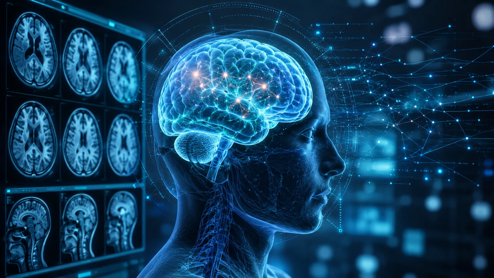
For decades, an Alzheimer’s diagnosis often felt like entering a room filled with unanswered questions. Doctors could identify memory loss, families could notice personality changes, and brain scans could reveal structural damage, but connecting those clues to a precise treatment plan remained difficult. Now, AI brain imaging for Alzheimer’s is beginning to change that experience by turning complex MRI and PET scans into clearer, more measurable clinical information. The technology is arriving at a crucial moment, as newer Alzheimer’s therapies require physicians to understand not only whether disease-related proteins are present, but also how the brain responds after treatment begins. Instead of serving only as a diagnostic snapshot, brain imaging is becoming an active guide throughout the care journey.
This shift is not built around a futuristic machine making medical decisions alone. It is happening through software that helps neurologists, radiologists, and nuclear medicine specialists examine patterns that can be difficult to measure consistently with the human eye. AI systems can map brain regions, calculate tissue volumes, quantify protein-related signals, and compare multiple scans taken months apart. They can also help highlight subtle abnormalities that deserve closer review before a physician prescribes or continues a therapy. In practical terms, the goal is not to replace medical expertise, but to give specialists a sharper visual dashboard for one of the most complicated diseases in modern healthcare.
Why Alzheimer’s Treatment Now Depends on Better Images
Alzheimer’s disease is not a single visible event that appears overnight. It develops through biological changes that may begin years before major memory symptoms become obvious, including the accumulation of amyloid plaques, the spread of tau-related damage, and the gradual loss of healthy brain tissue. Traditional cognitive testing remains important, but it cannot show exactly what is happening inside the brain. MRI can reveal structural changes, vascular conditions, bleeding, swelling, and patterns of atrophy, while PET imaging can help visualize disease-related molecular activity. When those images are analyzed together, they can create a more detailed picture of whether a patient may be eligible for treatment and how carefully that treatment should be monitored.
The need for accurate imaging has grown alongside the arrival of anti-amyloid therapies designed for people in the early stages of Alzheimer’s disease. These treatments aim to reduce amyloid plaque in the brain and may slow cognitive decline for some eligible patients, but they also require careful screening and follow-up. Physicians must confirm that the person’s symptoms and biological markers fit the treatment criteria, while also checking for conditions that could increase risk. Brain scans may be needed before treatment, during the early infusion period, and again if concerning symptoms appear. That creates a much heavier imaging workload than the older model of using a scan mainly to support an initial diagnosis.
The challenge is that every scan contains an enormous amount of visual data. A radiologist may need to inspect hundreds of image slices, compare different MRI sequences, review previous examinations, and evaluate tiny changes that could influence a treatment decision. Even highly trained specialists can face variation in how findings are measured or described, especially when hospitals use different scanners and workflows. AI-assisted analysis offers a way to standardize parts of that process without removing the physician from the final judgment. By automatically organizing and quantifying key findings, the software can help clinicians spend more time interpreting what those findings mean for the person sitting in front of them.
How AI Brain Imaging for Alzheimer’s Works
At the center of AI brain imaging for Alzheimer’s is a collection of machine learning models trained to recognize anatomical structures and clinically relevant patterns. After an MRI or PET scan is uploaded, the software can segment the brain into specific regions, such as the hippocampus, cerebral cortex, ventricles, and white matter. It may then calculate the volume of each region and compare the results with reference data from people of similar ages or clinical backgrounds. This allows doctors to see whether certain areas appear unusually small, asymmetric, or changed compared with an earlier scan. The final output usually includes visual overlays, quantitative measurements, and structured reports rather than a simple yes-or-no diagnosis.
PET imaging adds another layer because it can be used to evaluate the distribution of biomarkers associated with Alzheimer’s pathology. AI software can help calculate standardized uptake value ratios, often shortened to SUVR, which estimate how strongly a tracer signal appears in selected brain regions. A consistent automated calculation can make it easier to compare scans across time or evaluate whether a therapy is reducing amyloid burden. The system may also generate color-coded maps that translate subtle numerical differences into visual patterns physicians can review more quickly. These tools do not eliminate the need for expert interpretation, but they can make highly technical imaging data easier to navigate.
Longitudinal analysis is especially important because Alzheimer’s care is becoming less about a single appointment and more about tracking a biological process. An AI platform can align an older scan with a newer one, compensate for differences in positioning, and measure how specific structures or signals have changed. That capability matters when clinicians are trying to distinguish real disease progression from normal imaging variation. It can also help determine whether an apparent change is large enough to affect the treatment plan. Over time, the patient’s scan history begins to function like a visual timeline rather than a disconnected collection of medical images.
A New Tool for Monitoring Treatment Risks
One of the most urgent uses of AI-assisted imaging involves amyloid-related imaging abnormalities, commonly known as ARIA. These abnormalities can occur in some patients receiving anti-amyloid therapies and may involve brain swelling, fluid accumulation, small areas of bleeding, or deposits of blood-breakdown products. Many cases are mild or do not cause obvious symptoms, but some can become serious and require treatment to be paused or stopped. Because ARIA is identified primarily through MRI, imaging quality and timing are central to safe treatment. The growing number of patients entering therapy means healthcare systems need reliable ways to detect these findings early and manage them consistently.
AI tools may assist by highlighting regions that show possible edema, microhemorrhages, or other changes associated with ARIA. The software can compare a current MRI with the patient’s baseline examination and flag differences that might otherwise require lengthy manual review. It can also count or measure small lesions, helping specialists evaluate whether the pattern has changed enough to alter treatment. However, a highlighted region is not automatically a confirmed complication, and false alarms remain possible. The radiologist must still consider the imaging sequence, patient history, symptoms, medications, and treatment stage before reaching a conclusion.
This is where the conversation around medical AI becomes more grounded than the hype surrounding consumer image generators. In clinical imaging, speed matters, but reliability, traceability, and context matter even more. A model that produces a visually impressive result but cannot explain how it reached that result may be difficult to trust in a high-risk treatment setting. Hospitals need systems that preserve the original scans, display measurable findings, and allow clinicians to verify every automated suggestion. The strongest products will likely be those that behave less like mysterious prediction engines and more like transparent measurement assistants.
From Diagnosis Software to a Full Treatment Platform
The broader trend in medical imaging AI is a move away from single-purpose tools. Earlier software might have been built to detect one condition or calculate one measurement, but newer platforms aim to support multiple stages of the clinical pathway. In Alzheimer’s care, that can include screening, differential diagnosis, treatment eligibility, baseline documentation, safety monitoring, and assessment of treatment response. A unified system could combine structural MRI analysis, amyloid PET quantification, lesion tracking, and follow-up comparisons within one interface. That approach reflects how clinicians actually work, because treatment decisions rarely depend on a single image or metric.
This full-cycle model also explains why brain imaging companies are increasingly focused on workflow integration. A powerful algorithm has limited value if medical staff must manually export scans, rename files, switch between multiple applications, and rebuild reports from scratch. Hospitals need tools that connect with existing picture archiving systems, electronic medical records, and radiology reporting environments. Results must arrive quickly enough to support a real appointment rather than appearing days later in a separate research dashboard. Successful adoption will depend as much on usability and interoperability as on the accuracy of the underlying AI.
For patients and caregivers, smoother integration could reduce some of the confusion surrounding advanced Alzheimer’s treatment. The current care pathway may involve neurologists, imaging centers, infusion clinics, laboratory testing, genetic risk discussions, and repeated follow-up visits. When information is fragmented, families can struggle to understand why another scan is needed or what a new result means. A well-designed imaging platform could help care teams present changes more clearly by showing visual comparisons and standardized measurements. It cannot remove the emotional weight of the disease, but it may make the medical process feel less opaque.
Why This Moment Matters for Visual Technology
The emergence of Alzheimer’s imaging platforms shows how visual technology is expanding far beyond design, entertainment, and digital media. Computer vision is increasingly being used to interpret biological structures that are too detailed, subtle, or multidimensional for fast manual analysis. In this setting, an image is not merely something to look at; it is a dense layer of measurable information. AI can transform that information into maps, trends, comparisons, and alerts that support human reasoning. This makes healthcare one of the clearest examples of how visual innovation can move from impressive demonstrations to decisions with real-world consequences.
The technology also represents a shift from generative AI toward analytical AI. Generative systems create new images, videos, or text, while medical imaging platforms focus on interpreting evidence that already exists. Their value comes from precision, reproducibility, and the ability to find patterns across thousands of data points. That difference matters because public conversations about AI often treat every system as if it works the same way. In reality, an algorithm that segments a hippocampus or quantifies a PET tracer operates under a very different set of expectations than a tool that creates concept art from a prompt.
Visual interfaces will still play a major role in whether clinicians trust and use these systems. A report filled with unexplained percentages may be technically accurate but clinically frustrating. Doctors need to see where a measurement came from, how it compares with prior scans, and whether the model’s segmentation follows the actual anatomy. Clear overlays, adjustable views, confidence indicators, and side-by-side comparisons can make the AI’s output easier to verify. Good medical software design is therefore not decoration; it is part of the safety architecture.
The Human Impact Behind the Data
Behind every MRI series is a person who may already be dealing with uncertainty, fear, or a changing sense of independence. A patient being evaluated for early Alzheimer’s disease may still be working, driving, managing finances, and participating fully in family life. The decision to begin treatment can involve difficult tradeoffs between potential benefit, infusion schedules, side effects, costs, and the burden placed on caregivers. More detailed imaging can support that decision, but it should never turn the patient into a collection of colored brain regions. The best use of AI is to strengthen a thoughtful clinical conversation, not to replace it with a score.
Clearer measurements may also help families understand why a doctor recommends one path instead of another. For example, imaging might reveal that a person has significant vascular damage, a pattern of microbleeds, or another condition that changes the risk calculation. In other cases, PET analysis may support the presence of amyloid pathology and help confirm that a patient fits the biological profile studied in clinical trials. These findings can make treatment discussions more specific, but they do not guarantee a particular outcome. Alzheimer’s remains a highly individual disease, and two people with similar scans may experience different rates of decline.
There is also a risk that advanced imaging creates a new divide between patients who can access specialized care and those who cannot. Many communities already face long waits for neurologists, limited availability of PET scanning, high out-of-pocket costs, and difficulty traveling to infusion centers. AI may improve efficiency inside major hospitals while doing little for people who cannot reach those hospitals in the first place. Developers and healthcare leaders will need to think beyond algorithm performance and consider whether the technology can function across regional clinics, community hospitals, and lower-resource settings. A breakthrough that remains concentrated in a few elite centers will not transform Alzheimer’s care at the scale society needs.
What Hospitals Must Get Right
Before adopting an AI imaging system, hospitals need evidence that it performs reliably across different patient populations and scanner environments. A model trained mostly on data from one country, age group, or device manufacturer may not behave the same way elsewhere. Brain anatomy, vascular risk, disease presentation, and image quality can vary, creating opportunities for hidden bias. Clinical validation should therefore include diverse datasets and real-world conditions rather than only carefully selected research scans. Ongoing monitoring is also necessary because performance can change as hospital protocols, software versions, and patient populations evolve.
Healthcare organizations must also define responsibility when an AI suggestion conflicts with a physician’s interpretation. The answer cannot be to follow the algorithm automatically, nor should staff ignore it simply because it is new. Hospitals need protocols explaining when a flagged finding requires additional review, how disagreements are documented, and who makes the final decision. Training should include both the strengths and limitations of the system, including common failure cases. When clinicians understand how a tool can be wrong, they are better prepared to use it safely.
Data protection is another major issue because brain scans are deeply personal medical records. AI platforms may process large image files in local hospital servers, private cloud systems, or external computing environments. Each setup creates questions about encryption, access controls, retention policies, and whether patient data may be reused to improve future models. Consent and governance practices must be understandable rather than buried in technical language. Patients should not have to sacrifice privacy simply because their care involves advanced analysis.
What Patients and Caregivers Should Ask
Patients do not need to become imaging experts, but they can ask practical questions about how AI is being used in their care. One useful question is whether the software is measuring brain structures, analyzing PET biomarkers, monitoring treatment side effects, or performing several of those tasks. Another is whether a radiologist reviews and confirms every automated result before it influences a treatment decision. Families can also ask how a new scan compares with the baseline image and whether the observed difference is clinically meaningful. These questions shift the conversation away from vague claims about “smart technology” and toward the specific role the tool plays.
It is equally important to ask what the imaging result cannot reveal. A scan may support an Alzheimer’s diagnosis, but it cannot perfectly predict how quickly a person’s symptoms will progress. A reduction in amyloid signal does not necessarily mean that memory will return, and a stable MRI does not capture every aspect of daily function. Cognitive assessments, medical history, caregiver observations, and the patient’s own goals remain essential. AI imaging should be understood as one important piece of a much larger clinical picture.
Caregivers may also want a clear schedule for future scans and an explanation of symptoms that require urgent attention. Headache, confusion, dizziness, vision changes, weakness, nausea, or seizures can have many causes, but certain symptoms may require prompt evaluation during anti-amyloid treatment. The care team should explain whom to contact, where emergency imaging would be performed, and whether local clinicians have access to the treatment history. A sophisticated AI platform cannot compensate for a poorly communicated safety plan. Technology works best when it is supported by organized, responsive human care.
The Next Phase of AI-Powered Alzheimer’s Care
The next generation of brain imaging AI will likely combine more than MRI and PET data. Researchers are developing multimodal systems that can integrate blood biomarkers, cognitive scores, genetic information, medication history, and longitudinal imaging. In theory, these platforms could help estimate which patients are most likely to benefit from a therapy and which may face greater safety risks. They could also identify disease patterns that do not fit traditional categories, supporting more personalized treatment strategies. However, combining more data does not automatically create better care, especially if the model becomes too complex for clinicians to understand or validate.
Another likely development is greater automation of follow-up comparisons. Instead of requiring specialists to manually retrieve and align previous scans, software could automatically build a timeline of anatomical, molecular, and safety-related changes. Clinicians might receive a dashboard showing amyloid trends, brain volume changes, new lesions, and treatment milestones in one place. Such a system could make multidisciplinary meetings more efficient and reduce the chance that an important detail is buried in an older report. The value would come not from replacing specialists, but from organizing the growing amount of information they must manage.
AI may also help expand access by reducing the time required for expert image analysis. Smaller hospitals that lack dedicated neuroimaging specialists could use automated measurements to support consultations with regional medical centers. Cloud-based review networks might allow complex scans to be assessed remotely while keeping local physicians involved in care. Yet this model will require strong quality controls, dependable internet infrastructure, and clear lines of responsibility. Expanding access should never mean lowering the standard of review for patients outside major cities.
A Visual Turning Point in Alzheimer’s Treatment
The most important change is not that a computer can look at a brain scan. Software has assisted medical imaging for years, and automated measurements are not entirely new. What is different now is the role imaging plays in the emerging Alzheimer’s treatment pathway. Scans are becoming central to identifying eligible patients, establishing a baseline, watching for side effects, measuring biological response, and deciding whether therapy should continue. That expanded responsibility creates a strong case for tools that can make image analysis more consistent, transparent, and manageable.
Still, the future of Alzheimer’s care will not be determined by algorithm accuracy alone. Trust will depend on whether the technology works across diverse populations, integrates into real clinical workflows, protects sensitive data, and gives physicians results they can independently verify. Access will depend on whether healthcare systems can provide scans, specialists, infusions, and follow-up care beyond wealthy urban centers. Patients will judge the technology by whether it leads to clearer answers and safer decisions, not by how advanced the interface appears. The real test is whether innovation reduces uncertainty without creating new forms of inequality or confusion.
AI brain imaging for Alzheimer’s is therefore best understood as a bridge between visual data and more informed treatment. It can help reveal patterns hidden inside complex scans, track changes that unfold over time, and support the careful monitoring required by newer therapies. It cannot cure Alzheimer’s, predict every outcome, or replace the judgment of experienced clinicians. What it can do is give care teams a more precise view of a disease that has remained frustratingly difficult to see and manage. In a field where every treatment decision carries emotional and medical weight, that clearer view may become one of the most meaningful advances of the decade.